01-Jun-2026
For the heart to work properly, its electric signals and contractions must be timed together. The breakdown of this electrical-mechanical co-ordination can lead to various heart conditions, such as long QT syndrome.
Studying the normal function of the heart and how it goes wrong in certain disease states requires an understanding of how these electrical and mechanical signals are synchronised. However, this direct study has historically proved challenging for researchers due to incompatibility between the techniques used to study electrical and mechanical signals in heart cell and organoid models. Electrical signals are typically monitored using a Microelectrode Array (MEA), and mechanical signals using Atomic Force Microscopy (AFM). MEA gives information on the electrical signals of cells without being invasive. This is typically reported as a field potential (millivolts). AFM involves placing a cantilever with a fine tip over the sample of interest. As the tip of the cantilever interacts with the sample, movement of the cantilever and the force placed on it by the sample is measured. In terms of cell biology, this can give information on the mechanical properties of the cell such as action forces, beat rate, and elasticity. So far, researchers have struggled to collect electrical and mechanical data at the same time from the same sample.
However, researchers based at the CEITEC, part of the Czech Infrastructure for Integrative Structural Biology of Instruct-CZ, have recently developed an experimental approach and accompanying data analysis pipeline to allow the simultaneous measurement of electrical and mechanical signals from cell and organoid-based models of heart function.
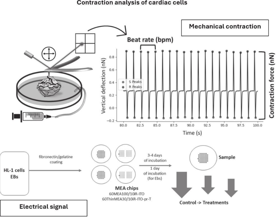
Figure 1 – Overview of Atomic Force Microscopy and Microelectrode Array methods for collecting mechanical and electrical data, respectively.
The team of scientists developed a way to capture and combine electrical and mechanical information, to allow for a more holistic study of how heartbeats are controlled. The system involves an experimental setup in which heart organoid models (called embryoid bodies) are measured with both MEA and AFM simultaneously. The electrical and mechanical data are then fed into an analysis pipeline built on python scripts developed by the team, called “CardioScripts”. As well as combining the electrical and mechanical data, CardioScripts can automatically identify the various parts of the waveform commonly used to represent electrical heart signals, called the QRST complex.
The team showed that their system worked by looking at the electro-mechanical synchronisation of embryoid bodies under normal and drug-treatment conditions. Without treatment, embryoid bodies showed synchronised behaviour with an electrical signal closely followed by contraction - as happens in the healthy human heart. However, when embryoid bodies were treated with the drug lidocaine, the electrical and mechanical signals became de-coupled.
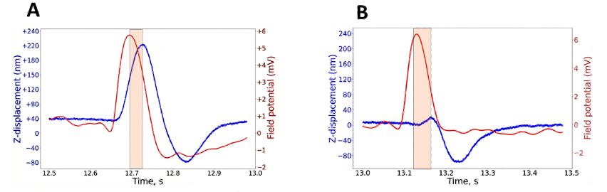
Figure 2 – Electrical and mechanical coupling as captured by the MEA-AFM integrated setup. Representative curves showing electrical (red) and mechanical (blue) signals are (a) coupled under normal conditions and (b) de-coupled by treatment with lidocaine.
"What makes this work important is that it brings together two complementary views of cardiomyocyte function — the electrical signal and the mechanical response. In our field, that is essential for understanding how cardiac cells behave in health, how they respond to drugs, and how electromechanical coupling begins to fail in disease. This study builds on our long-term focus on AFM-based biomechanical characterization of cardiac models and extends it toward a more integrated and reproducible framework.
The work was made possible by the combination of expertise and instrumentation available through the Nanobiotechnology Core Facility at CEITEC Masaryk University, which provides advanced AFM, multielectrode array recording, and correlated biophysical measurements for living cells and biomolecular systems. As part of CIISB / Instruct-ERIC, the facility gives researchers access to high-end infrastructure and technical support that make this kind of interdisciplinary method development feasible.”
Says Jan Přibyl, who runs the Nanobiotechnology Core Facility at CEITEC Masaryk University, and who headed the team of scientists that produced CardioScripts.
The team also included Daniil A. Kabanov, Simon Vrana, Deborah Beckerová, Vladimir Rotrekl, and Martin Pesl, and represents a collaboration between Masaryk University and St Anne’s University hospital, both based in Brno, Czech Republic.
You can read the full paper “A comprehensive system of algorithms for characterization of cardiomyocyte mechanical and electrical function”, published in Biomedical Signal Processing and Control here. Additionally, all scripts developed for the paper are available for free on GitHub, and the accompanying datasets are freely available on Zenodo.
This work was supported by the Czech Infrastructure for Integrative Structural Biology at the Instruct-CZ centre.
